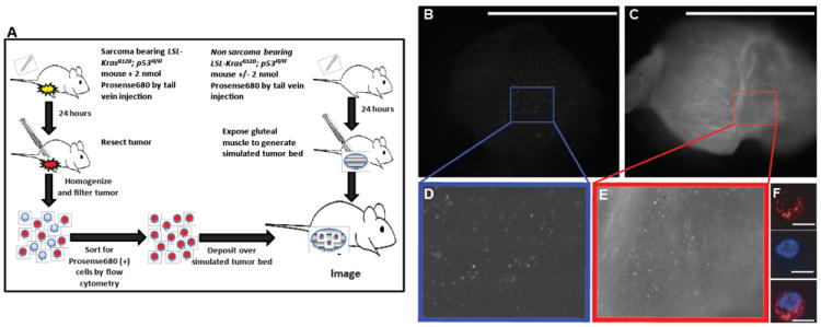
Figure 4.

Imaging individual cells transferred into simulated tumor beds in vivo. (A) Cells were obtained by flow cytometry from a sarcoma bearing mouse 24 hours after Prosense 680 injection. Individual Prosense (+) cells from the sarcoma were detected by the hand-held imaging device when placed in a simulated tumor bed from a mouse not injected (B) and injected (C) with Prosense 680. (F) Prosense signal was confirmed to originate from individual cells by confocal microscopy in the Prosense channel (top, red) and nuclear Hoechst stain (middle, blue), which were merged together (bottom). Insets in A and B show a 2.5x magnification of the highlighted area (D, E). Scale bars: 5 mm for A, B; 10 μm for D, E.
