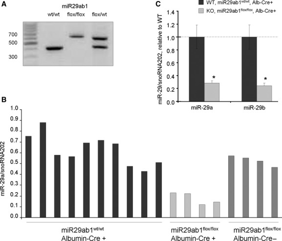
Fig. 2.

miR-29 expression in vivo. (A) PCR analysis of DNA from mouse tail. Flox sites were inserted upstream and downstream of miR29ab1 site. In presence of Cre, Flox sites could be digested and miR29ab1 site cut-off. Cre was expressed under the albumin promoter and therefore miR-29a and miR-29b was selectively knocked out in hepatocytes. (B) RNA was isolated from liver tissues from wild-type (miR29ab1wt/wt, Cre+), knockout (miR29ab1flox/flox, Cre+) or floxed mice (miR29ab1flox/flox, Cre−), and miR-29a expression was assessed using a Taqman based quantitative RT-PCR assay. Mir-29a expression was selectively reduced in miR29ab1flox/flox, Cre+ animals. (C) The expression of miR-29a and miR-29b was assessed by qRT-PCR and normalized to that of smoRNA202. The bars represent mean and standard error of expression in four mice, relative to wild-type controls (WT). *P < 0.05 relative to controls.
