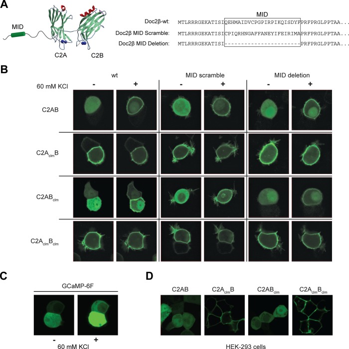
FIGURE 4:
Constitutive plasma membrane localization of mutant forms of Doc2β. (A) The wt and Ca2+-ligand mutant forms of Doc2β were modified by deleting or scrambling the MID. (B) These constructs were fused to GFP, transfected into PC12 cells, and imaged using confocal microscopy before and after depolarization with 60 mM KCl. (C) Increases in [Ca2+]i were monitored using the high-affinity Ca2+-sensor GCaMP-6F. GCaMP-6F was expressed in PC12 cells, and the fluorescence intensity was monitored before and after depolarization with KCl. (D) Live-cell images of HEK 293 cells expressing the indicated GFP-Doc2β fusion protein.

