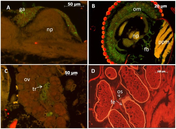
Figure 2. Infection of various Aedes aegypti tissues with rRVF-wt.
A) ventral nerve cord, showing infection of the ganglia B) head, showing infection of the ommatidia and fat body, C) ovary, showing infection of the tracheal cells D) ovarioles, showing infection of the follicular epithelium and ovariole sheath. Abbreviations: fb = fat body; fe = follicular epithelium; ga = thoracic ganglia; np = neuropile; om = ommatidia; ov = ovary; os = ovariole sheath; ppm = pharyngeal pump musculature; tr = tracheoles.

