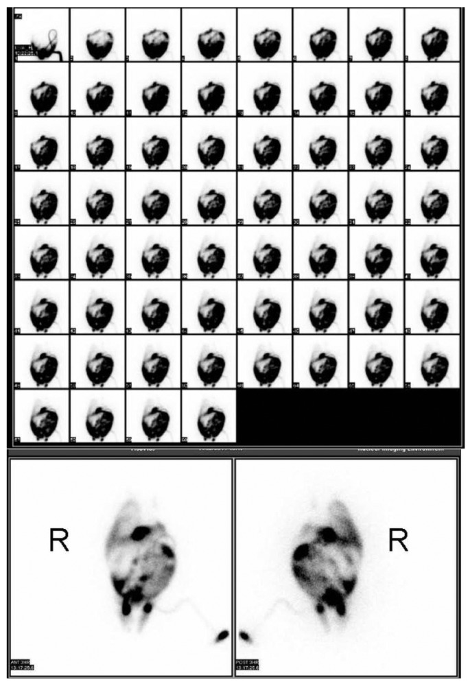
Figure 4 —

Peritoneal scintigraphy demonstrates leakage of peritoneal fluid into the right pleural cavity and also minimal drainage into the left pleural space. Early images (top) were taken at 1-minute intervals. The later images were taken at 3 hours after infusion. R = patient’s right side.
