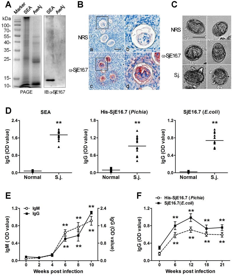
Figure 2. Localization and immunogenicity of SjE16.7.
(A) Expression of SjE16.7 in the egg stage of S. japonicum. Extracts obtained from adult and egg stages of S. japonicum were separated by SDS-PAGE and blotted with anti-SjE16.7 sera. SEA, soluble egg antigen. AwAj, adult worm antigen of S. japonicum. (B) Immunohistological detection of SjE16.7 in S. japonicum eggs. Sections of livers from S. japonicum-infected mice were incubated with normal rabbit sera (a,b) or rabbit anti-SjE16.7 sera (c, d) NRS, normal rabbit sera; α-SjE16.7, rabbit anti-SjE16.7 sera. Right graphs are better seen in the magnification of the insets of upper graphs. Original magnification ×40, scale bar = 50 µm. (C) Precipitate formation around live eggs cultured in vitro with anti-SjE16.7 sera (α-SjE16.7) and S. japonicum infected rabbit sera (S.j.), but not normal rabbit sera (NRS). Original magnification ×40, scale bar = 50 µm. (D) ELISA detection of anit-SEA or SjE16.7 IgG in S. japonicum infected mice. The sera were collected before (Normal) and 42 days after S. japonicum infection (S.j.). SEA, soluble egg antigen; His-SjE16.7, recombinant His-SjE16.7 protein from Pichia yeast; SjE16.7, recombinant SjE16.7 (without GST tag) protein from E. coli. ** P<0.01. (E, F) Time course determined by ELISA. Sera from S. japonicum infected mice (E) or rabbits (F) were collected at different time points. Anti-His-SjE16.7 IgG or IgM antibodies were measured by ELISA. Each group consisted of at least 5 animals. Data are presented as optical density at 450 nm (mean ± SD). ** P<0.01 vs. zero time point.

