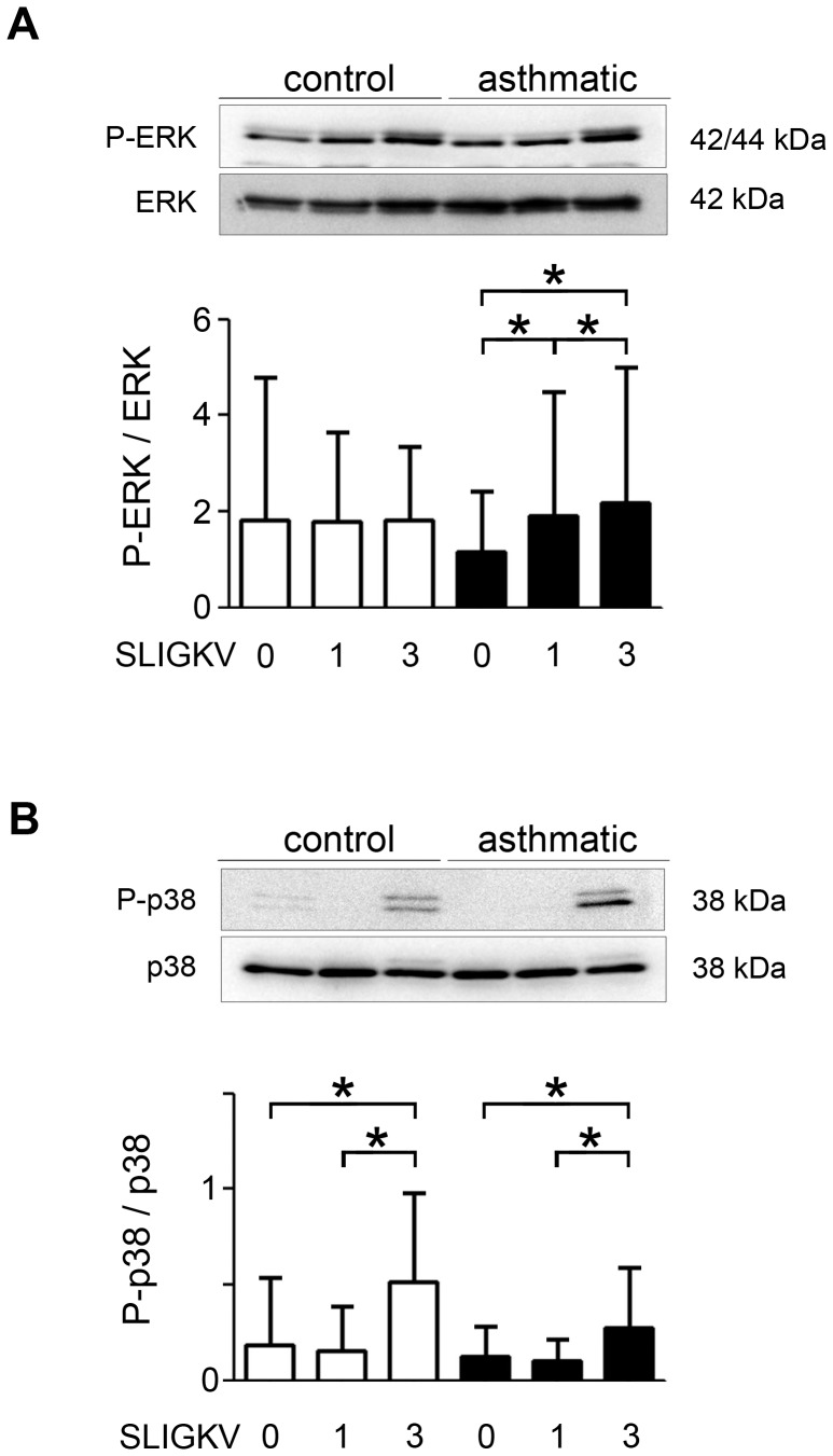
Figure 5. Increased asthmatic bronchial smooth muscle cell phosphorylation of ERK and p38 following repeated PAR-2 stimulations.
Phosphorylation of ERK (A) and p38 (B) was measured using western blot following stimulation for 0, 1 or 3 days by 10−4 M SLIGKV-NH2. Representative blots stained with anti-Phospho-ERK (P-ERK), anti–ERK, anti-Phospho-p38 (P-p38) and anti-p38 antibodies are shown. Bronchial smooth muscle cells were obtained from asthmatic (black bars, n = 8) and control subjects (white bars, n = 7). Results are expressed as mean ± SD. *P<0.05 using paired Wilcoxon-rank tests.

