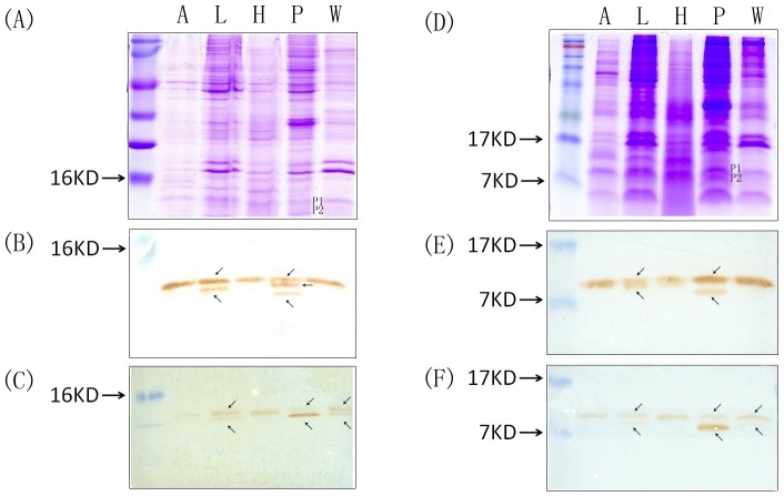
Figure 4. Functional expression of CSP-RDDs.
Electrophoretic separation and Western blot analysis of CSP proteins in the antennae (A), legs (L), heads (H), pheromone gland (P) and wings (W) of female B. mori. Protein extracts corresponding to 10 antennae, 30 legs, 5 heads, 5 pheromone glands and 20 wings equivalent were subjected to 15% SDS-PAGE (A. and D.) and transferred to nitrocellulose membranes (B–C. and E.F.). Nitrocellulose blots were labeled with two different antisera: “anti-CSP1” (B. and E.) and “antiCSP14” (C. and F.). Positions of molecular weight markers are indicated on the left of the gel. Proteins of 9 to 14 kDa are labeled with the two CSP antibodies in the pheromone gland, legs and wings samples.

