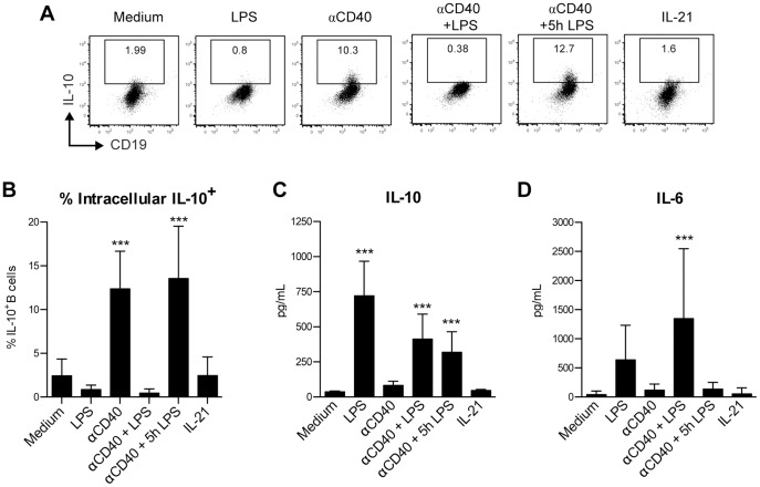
Figure 2. PerC B-1a cells secrete IL-10 and IL-6 similarly to the undifferentiated PerC B cell population.
FACS-purified CD19+ CD11b+ CD5+ PerC B-1a cells were cultured for 48 h in the presence of medium or indicated stimuli. Intracellular IL-10 staining was performed after additional stimulation with PMA/ionomycin/LPS and both representative flow cytometric plots (A) and the percentage of IL-10+ B cells (B) are shown, whereas the levels of IL-10 (C) and IL-6 (D) were determined directly from the supernatant (before restimulation took place) using Luminex. Shown in B-D are the mean ± S.D. of pooled results from two identical replicate experiments (with 2–5 technical replicates each) that produced similar data. Statistically significant differences between the various stimuli and the medium control (B-D) of PerC B-1a cells were calculated using a one-way ANOVA with Bonferroni post-hoc test. ** p<0.01, *** p<0.001.

