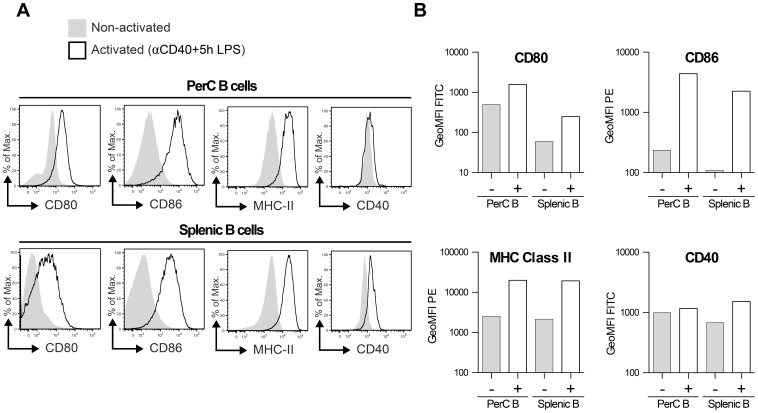
Figure 3. PerC– and Splenic B cells possess different surface markers after activation.
Indicated B cell populations were stained extracellular with mAbs against CD19 and indicated markers after both a 48 h period of stimulation using αCD40+5 hr LPS (+: activated; see materials and methods) and after being isolated freshly (–: non-activated). Shown in A are expression levels of indicated markers on CD19+ B cells (representative plots of two identical replicate experiments), in B the average geometric mean fluorescence intensity (GeoMFI) levels from two identical replicate experiments.

