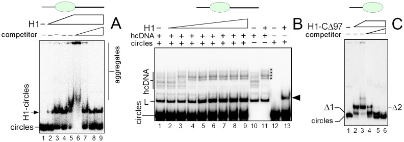
Figure 5. Binding of histone H1 to DNA minicircles.
(A) Titration of DNA minicircles with histone H1. 32P-labeled DNA minicircles of 66-bp (∼30 pM) were titrated with histone H1 (2, 4, 8 and 15 nM, lanes 2–5) in the absence of competitor DNA. The H1-minicircles complex (prepared at 15 nM H1, lane 5) was also titrated with increasing amount competitor λ-DNA (10, 102, 103 and 104-fold mass excess of unlabeled competitor DNA over 32P-labeled minicircles; lanes 6–9, left to right). L, linear DNA of 66-bp. (B) Competition of DNA minicircles for histone H1 binding to hcDNA. An equimolar mixture of 32P-labeled DNA minicircles (66-bp) and hcDNA (∼30 pM) was titrated with histone H1 (2, 6, 9, 12, 18, 30, 50, 80 nM, lanes 2–9). 32P-labeled hcDNA without (lane 10) or with (lane 11) 15 nM H1 (the H1-hcDNA complexes are indicated by asterisks). 32P-labeled DNA minicircles without (lane 12) or with (lane 13) 15 nM H1. Arrowhead indicates position of the H1-DNA minicircles complex. (C) Binding of H1-CΔ197 to DNA minicircles. 32P-labeled DNA minicircles (∼30 pM) were titrated with increasing amounts of histone H1 lacking the C-terminal domain (peptide H1-CΔ97) (10 and 20 nM, lanes 2 and 3) in the absence of competitor DNA. Complex (H1-CΔ97)-DNA minicircles (prepared at 20 nM H1 peptide, lane 3) was also titrated with increasing amounts of competitor λ-DNA (103, 104 and 105-fold mass excess of unlabeled competitor DNA over 32P-labeled DNA minicircles, lanes 4–6, left to right). Δ1 and Δ2 indicate the (H1-CΔ97)-DNA minicircles complexes. H1-DNA complexes were resolved on 8% or 6% polyacrylamide gels in 0.5×TBE and visualized by autoradiography as detailed in the “Materials and Methods”.

