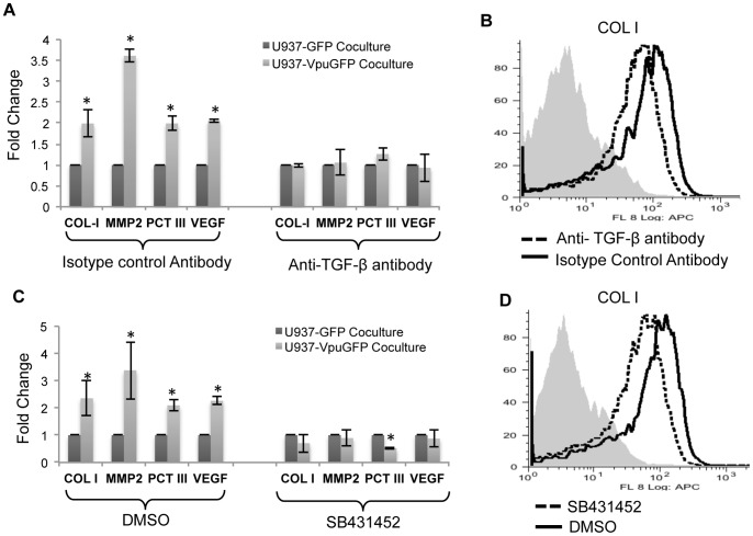
Figure 4. TGF-β secreted from Vpu-expressing monocytic cells is responsible for the profibrogenic effects on LX2 stellate cells.
(A) LX2 cells were cocultured with either U937-GFP or U937-VpuGFP cells together with either a pantropic anti-TGFβ antibody or an isotype control antibody as described in Methods. Quantitative RT-PCR was then carried out to estimate the expression levels of mRNAs for COL-1, αSMA-1, PCT-III, MMP2 and VEGF. All expression levels are shown as fold changes normalized to the U937-GFP control. (B) LX2 cells were cocultured with U1-scrambled cells together with either a pantropic anti-TGFβ antibody or an isotype control antibody, and flow cytometry was used to estimate the expression levels of intracellular COL-1. (C) LX2 cells were cocultured with either U937-GFP or U937-VpuGFP cells together with the TGF-β receptor inhibitor SB431452 (or DMSO as control) as described in Methods. Quantitative RT-PCR was then carried out to estimate the expression levels of mRNAs for COL-1, MMP2, PCT-III, and VEGF. All expression levels are shown as fold changes normalized to the U937-GFP control. (D) LX2 cells were cocultured with U1-scrambled cells in the presence of either SB431452 or DMSO, and flow cytometry was used to estimate the expression levels of intracellular COL-1. Error bars represent mean ± SD from three independent experiments; *p<0.05.

