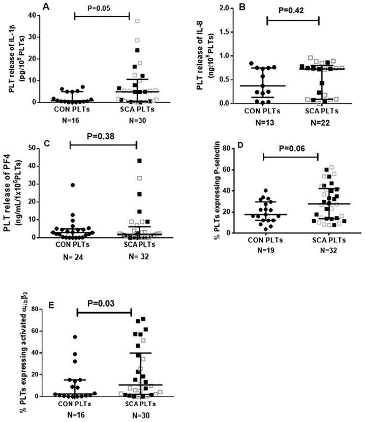
Figure 1. Platelets as inflammatory cells in SCA.
Release of (A) IL-1β, (B) IL-8, (C) platelet factor 4 (PF4) from platelets of healthy control individuals (CON) and SCA patients in steady state (SCA). Cytokine release was determined by ELISA in platelet suspensions (1×10⁸ PLT/ml) after incubation for 4 h (37°C, 5% CO2). Expressions of (D) P-selectin and (E) activated αIIbβ3 integrin on the surface of platelets from healthy control individuals (CON) and SCA patients in steady state (SCA). Medians and interquartile ranges are depicted. Adhesion molecule expression was determined by flow cytometry. Some of the data included in the data sets depicted in graphs 1D and 1E have been previously published in table form [14]. Closed black square symbols represent SCA patients not on HU therapy; open grey square symbols represent patients on HU (15–30 mg/kg/day) therapy.

