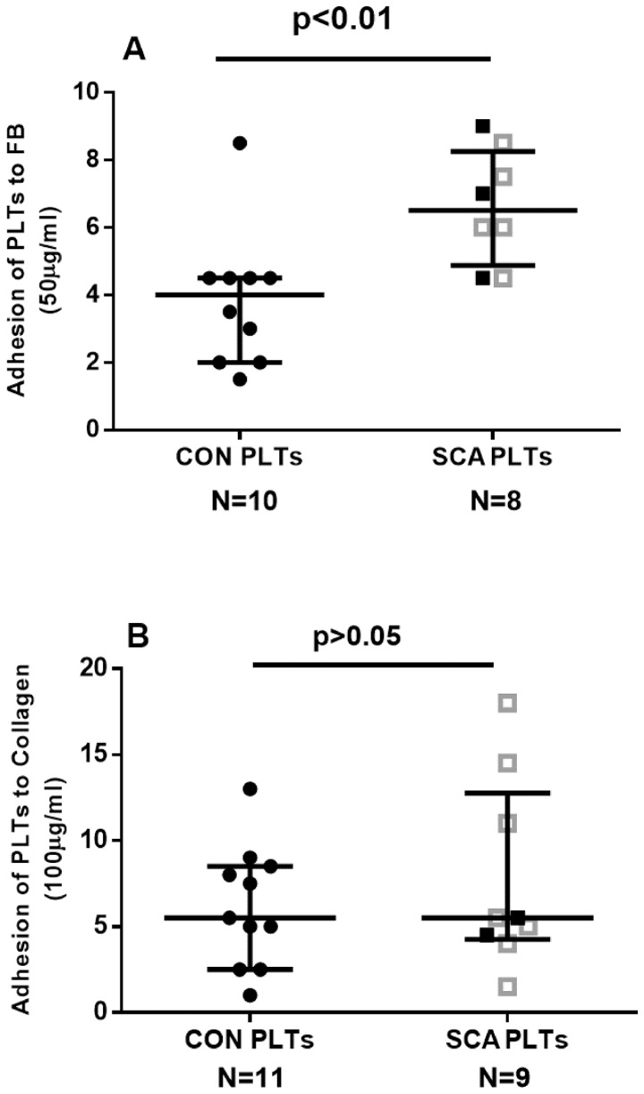
Figure 2. Adhesive properties of platelets.
A microfluidic assay was used to determine the adhesion of platelets from healthy control individuals (CON) and SCA patients in steady state (SCA) to (A) 50 µg/ml fibrinogen and (B) 100 µg/ml collagen. Platelets (1×10⁷ PLTs/mL) were perfused over microchannels (400 µm width) coated with immobilized ligands at a shear stress of 0.3 dyne/cm2 for 3 min at 37°C; adhesion was detected using brightfield inverted microscopy. The adhesion of platelets to duplicate microchannels was recorded at three positions in each channel and analyzed using the DucoCell analysis program, recording the mean number of platelets adhered to an area of 0.08 mm2. Medians and interquartile ranges are depicted. Closed black square symbols represent SCA patients not on HU therapy; open grey square symbols represent patients on HU (15–30 mg/kg/day) therapy.

