
Figure 1.
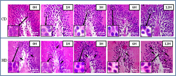
Representative photographs showing HD-induced histological changes in DG region of hippocampus. HD induced shrinkage and scattering of neuronal cells with gradual decrease in number during early period (Figs. HD, 1–6 H; arrows) as compare to their respective CD groups (Figs. CD, 1–6 H). CD induced disruption of histoarchitecture of DG region and increased necrotic changes after 6 h (CD, 12 H). Enlarge view of morphological changes in neuronal cells of the same figure (inset). Scale bar = 50 μM.