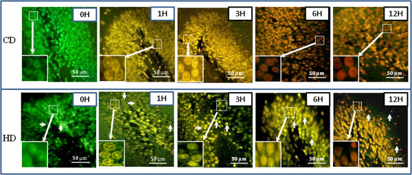
Figure 6.

Representative photographs showing HD induced apoptosis/necrosis in neuronal cells of the DG region of hippocampus. Initiation of apoptosis was observed after 1 h (Fig. HD, 1 H) as evidenced by greenish yellow color fluorescence, apoptosis after 3 h as evidenced by yellowish orange color fluorescence, which was found maximum after 6 h post- HD (Fig. HD, 6H; arrows) followed by necrosis as evidenced by orange-red or reddish color fluorescence (Fig. HD, 12 H; arrows). CD induced necrosis as early as 3 h and continued till the end of the experimental period (Figs. CD, 3-12H) as evidenced by orange-red or reddish color fluorescence. Enlarge view of apoptotic or necrotic changes in neuronal cells of the same figure (inset). Scale bar = 50 μM.
