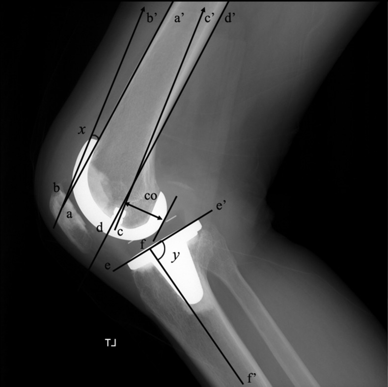
Fig. 2.
A lateral radiograph of the right knee shows the measurement of the sagittal alignment of the femoral and tibial components (x = sagittal femoral angle, and y = sagittal tibial angle). The posterior femoral condylar offset (CO) was evaluated by measuring the maximum thickness of the posterior condyle projected posteriorly to the tangent of the posterior cortex of the femoral shaft

