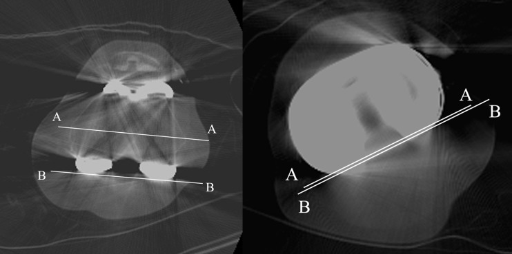
Fig. 3.
CT scan shows measurement of axial rotation of the femoral component in relation to the transepicondylar axis (A–A) and posterior femoral condylar line. CT scan shows measurement of axial rotation of the tibial component in relation to the posterior margins of the tibial plateau (A–A) and the tibial bearing (B–B)

