
Fig. 1.
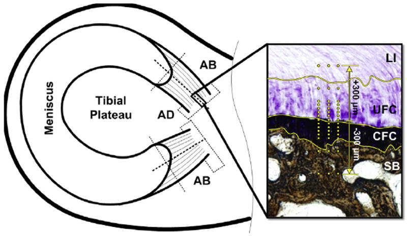
Schematic of sample location. AD sections used for histomorphometry. AB sections used for indentation and μCT. Inset – Meniscal enthesis stained with TB/VK to highlight the four unique regions: LI, UFC, CFC, and SB. Yellow lines highlight the demarcations between zones. Yellow dots represent location of indentation test points.