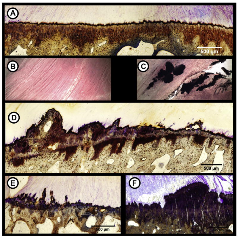
Fig. 2.

Pathophysiology in meniscal entheses due to OA. Healthy entheses have a smooth, intact TM (A) and are free from calcium deposits within the LI portion (B). Osteoarthritic meniscal entheses exhibited degenerative signs similar to other entheses such as calcium deposits within the LI zone (C), double TM formation (D), osteophyte formation (E), and microcracks/fissuring (F).
