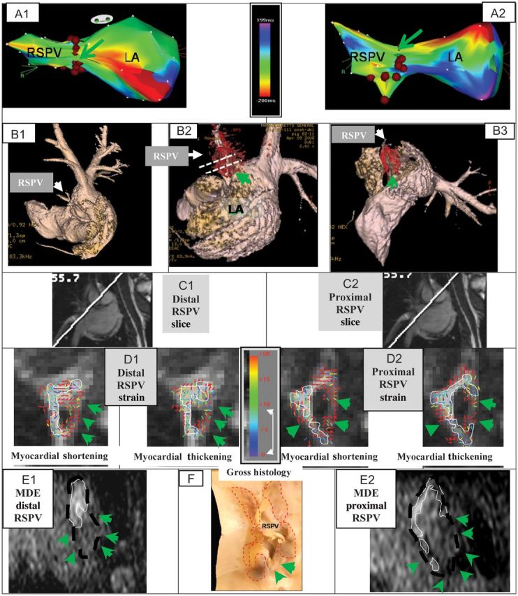
Figure 3.

Incomplete RF ablation of two swine RSPV. The first swine includes figures A–F, the second swine includes figures G–L. (A) EAM maps overlaid with RFA points (brown dots); Anterior (A1) and Superior (A2) views, showing gaps (green arrows). Actual size of gaps could not be determined from EAM. Activation maps were obtained prior to ablation. (B) Atrial luminal renderings (pink), segmented from 3D-MRA, are overlaid with a 3D-MDE scar map (red), showing the gaps in RSPV ablation (B2 and B3). (C) Images detailing the orientation of distal (C1) and proximal (C2) DENSE slices placed perpendicular to the RSPV ostium. Slice positions are also shown in 5B2. (D) Colour-coded DENSE myocardial shortening (negative sign) and myocardial-thickening (positive sign) strain magnitude for the distal (D1) and proximal (D2) slices. White lines outline regions with <8% strain. (E) Reformatted 3D-MDE slices at the position and orientation of D1 (E1) and D2 (E2). Dashed black line indicates position of RSPV wall, white lines outline enhanced ablated regions, and green arrows denote lack of enhancement (ablation gap). (F) Stained gross histology photograph of the LA wall at the RSPV ostium, overlaid with the areas (dashed red line) where transmural ablation lesions were found, with unstained ablation gaps denoted (green arrows). Incomplete RF ablation of the RSPV in another swine. (G) EAM maps overlaid with RFA points (brown dots); Anterior (G1) and Superior (G2) views, showing gaps (green arrows). Activation maps were obtained prior to ablation. (H) Luminal anatomy overlaid (H1–H4), with 3D-MDE scar, showing regions of incomplete ablation. Dashed white lines (H2 and H4) designate positions of 2D-DENSE slices placed perpendicular and parallel to RSPV ostium. Colour-coded DENSE strain for the distal (I1) and proximal (I2) perpendicular slices. White lines outline, 8% strain regions. (J) Position of ROIs placed on DENSE slice oriented along the RSPV to RIPV plane (see H4); (J1) ROI (red) encompassing the RSPV, and (J2) ROI encompassing the RIPV. (K) Strain results for ROIs covering RSPV (K1) and RIPV (K2). (L) Gross histology photograph of the LA wall at the RSPV ostium region, overlaid with ablation lesion regions (dashed red line).
