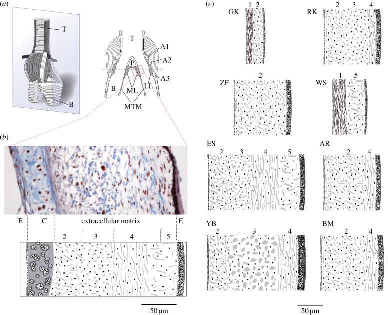
Figure 1.
(a) The syrinx consists of a cartilaginous framework, oscillatory soft tissues (labia) and muscles for controlling airflow and labial tension. The oscine syrinx contains two sound sources, each consisting of a pair of labia (lateral, LL and medial labium, ML) that are located near the tracheo-bronchial junction. T, trachea; A1–A3, syringeal cartilages; P, pessulus; B, bronchus, MTM, medial tympaniform membrane; ML, medial labium; LL, lateral labium. The grey box indicates the plane for sectioning. (b) Section of a starling medial labium illustrating its cellular and extracellular composition through trichrome stain. Thin epithelia (E) on each side encompass extracellular matrix, composed of collagen (blue) and elastin fibres (black). In this section, a part of the cartilage is also included (A1). The layer structure of the extracellular matrix is shown in a schematic below, identifying layers according to content and orientation of collagen fibres. Collagen fibres are oriented mostly randomly in layers 2 and 5, dorsoventrally in layer 3 and cranio-caudally in layer 4. Layer numbers (2–4) are used to identify similar organization in different species. Starlings do not have a layer dominated by elastin (layer 1 in (c)). (c) Schematics of composition of extracellular matrix and size differences of the medial labia in the eight songbird species. Layers are indentified by numbers corresponding to different fibre composition and orientation. These schemata reflect sections that did not contain cartilage. The layer thickness was drawn to scale and corresponds to the mean from two to eight specimens. Species GK, golden-crowned kinglet (Regulus satrapa); RK, ruby-crowned kinglet (Regulus calendula); ZF, zebra finch; WS, white-crowned sparrow (Zonotrichia leucophrys); ES, European starling (Sturnus vulgaris); AR, American robin (Turdus migratorius); YB, yellow-headed blackbird (Xanthocephalus xanthocephalus); BM, black-billed magpie (Pica hudsonia).

