Figure 2.
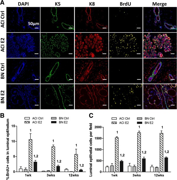
Rat strain-specific effects of 17β-estradiol on mammary epithelial cell proliferation. A, Representative fluorescent images of mammary tissues from ACI and BN rats, either sham treated (Ctrl) or treated with E2 for 1 week (n = 3). Column 1, nuclei identified by staining DNA with 4′,6-diamidino-2-phenylindole (DAPI, blue). Column 2, basal epithelial cells were identified by immunostaining for cytokeratin 5 (K5, green). Column 3, luminal epithelial cells were identified by immunostaining for cytokeratin 8 (K8, red). Colum 4, cells transiting S phase were identified by immunostaining for BrdU (yellow). Column 5, merged images from columns 1 through 4. Scale bars, 50 μm. B, The number of luminal epithelial cells (K8 positive) in S phase (BrdU positive) was quantified using a VectraTM multispectral fluorescence imaging system and illustrated as the percentage of total luminal epithelial cells. C, The number of luminal epithelial cells per field was quantified as an indicator of epithelial density. Each data bar in Panels B and C represents the mean ± standard error of the mean (SEM, n = 3). 1, p < 0.05 for comparison of E2 treated vs. sham treated rats of same strain. 2, p < 0.05 for comparison of E2 treated BN vs. treated ACI rats.

