
Fig. 5.
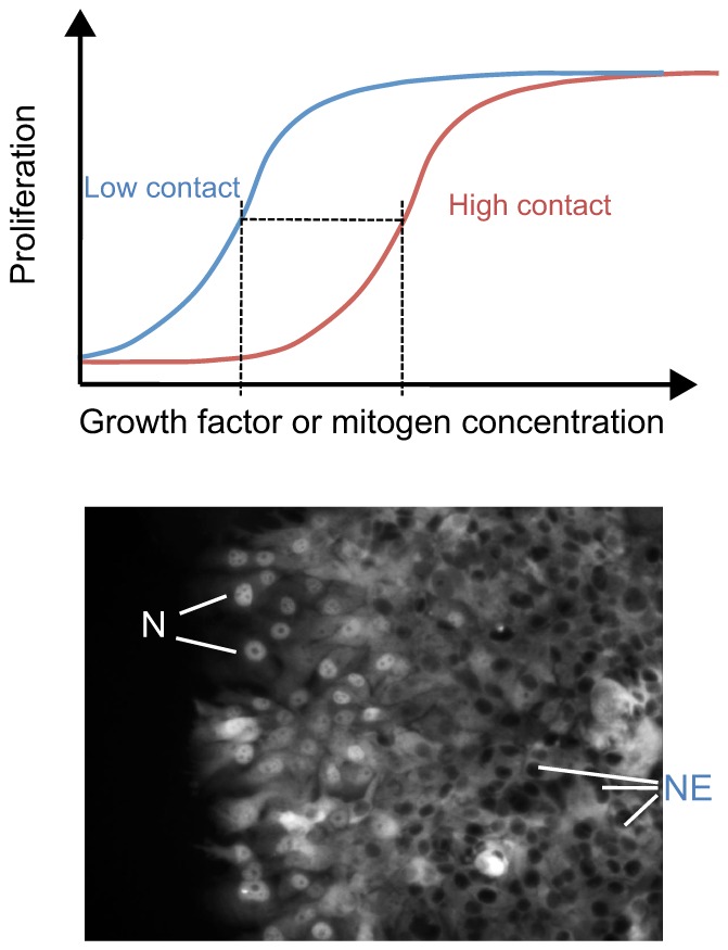
Role of contact inhibition and Hippo-YAP signaling in the spatial control of mitogenic signaling by growth factors. (Top) The graph shows the dose-response curves for growth factor-stimulated proliferation. A high degree of cell-cell contact shifts the curve on the x-axis, such that a higher concentration of growth factors is required to elicit the same response. This graph is an interpretation of the findings of Kim et al., 2009. (Bottom) Illustration how cell-cell contact regulates transcription factor activity through the Hippo pathway. At high cell density in the middle of the cell monolayer, the Hippo pathway is active, leading to nuclear exclusion (NE) of YAP and/or TEAD (TEAD is labeled by immunofluorescence staining in this example). At the edge of the culture where cells have lost contact inhibition, YAP and/or TEAD (as shown here) accumulate in the nuclei (N) and stimulate proliferation. Thus, proliferation is spatially regulated despite uniform levels of growth factors acting on these cells.