
Fig. 3.
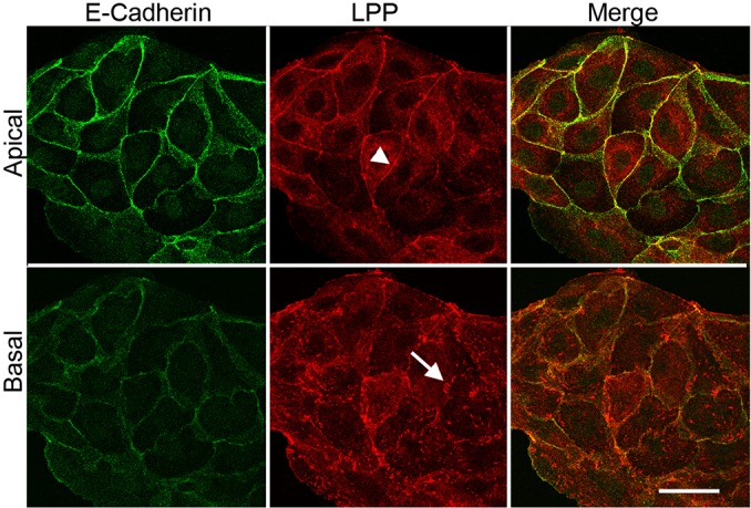
Immunofluorescent localization of LPP to cell contacts and focal adhesions. Top panels are confocal microscopic sections of the apical region of MDCK cells, which reveal that LPP (middle panel, red) is concentrated at cell contacts (white arrowhead) with E-cadherin (left panel, green and merge, right panel, yellow). Bottom panels are basal sections of the same cells, showing that LPP (middle panel, red) but not E-cadherin (left panel, green) is also localized to focal adhesions (white arrow). Scale bar: 20 µm.