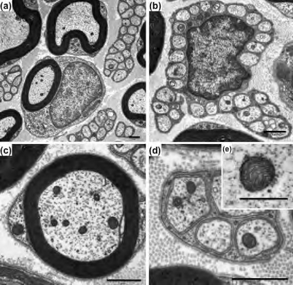
Figure 2.
Transmission electron micrographs of myelinated and unmyelinated fibers of the saphenous nerve from WT mice. Cross-section of myelinated and unmyelinated fibers (a); cross-section of unmyelinated fibers with Schwann cell nucleus (b); high magnification of myelinated (c) and unmyelinated (d) fibers; representative photograph of a normal axonal mitochondrion (e). Scale bar: 1 μm (a-d) and 0.5 μm (e).

