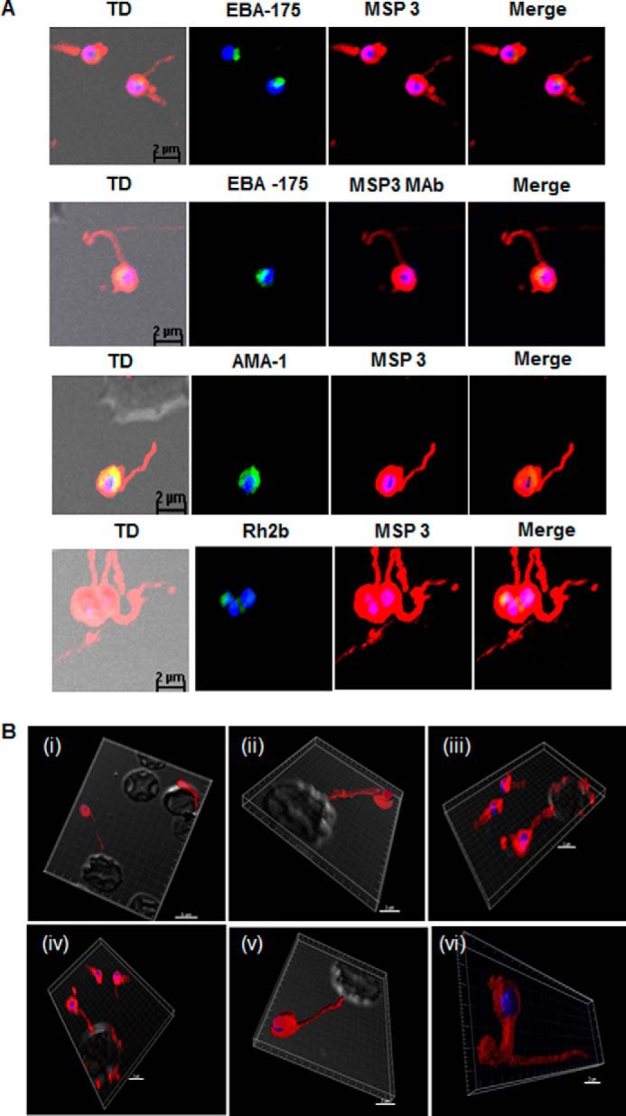
FIGURE 3.

Detection of fibrillar structures in P. falciparum merozoites by immunofluorescence assay. A, co-immunostaining of free merozoites with anti-MSP3 Abs along with Abs against EBA-175, monoclonal MSP3 Abs (MSP3 MAb), AMA-1, Rh2b. and MSP5. In all the slides. long fibrous structure attached to free merozoite was detected by anti-MSP3 Abs. Monoclonal MSP3 antibodies also stained these fibrillar structures associated with merozoites, but other marker protein did not detect fibrillar assemblies. TD, transmission differential interference contrast/ B, association of fibril-like assemblies with erythrocytes. Cytochalasin D-treated merozoites when incubated with fresh RBC showed the staining of the fibril-like structures possessing MSP3 associated with merozoites and RBC membrane. Images from panels i–v are the various fields depicting similar association. Panel vi shows single free merozoite with long fibril-like structure stained with anti-MSP3 Ab.
