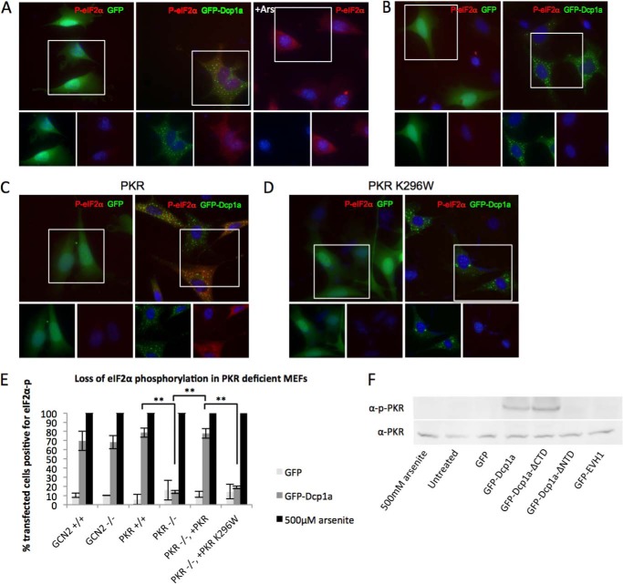
FIGURE 5.
PKR is activated by GFP-Dcp1a expression. A, WT MEFs electroporated with 1 μg/1.5 × 105 cells of pcDNA4-GFP (left panel) and peGFP-Dcp1a (center panel) or treated with 500 μm sodium arsenite (Ars) for 30 min (right panel). B, PKR-deficient (−/−) MEFs electroporated with 1 μg/1.5 × 105 cells of pcDNA4-GFP (left panel) and peGFP-Dcp1a (right panel). PKR−/− MEFs were coelectroporated with 1 μg/1.5 × 105 cells of pcDNA4-GFP or peGFP-Dcp1a and wild-type PKR (C) or inactive PKR K296W (D). Each set of MEFs was incubated with an antibody against phospho-eIF2α (S51) (red). E, quantification of eIF2α phosphorylation induced in electroporated cells in response to GFP or GFP-Dcp1a expression in several MEF cell lines as described under “Experimental Procedures.” **, p < 0.0001. Error bars indicate mean ± S.E. F, immunoblot of A549 cells transfected with the indicated plasmids and corresponding levels of total and phospho-PKR (Thr-446).

