
FIGURE 5.
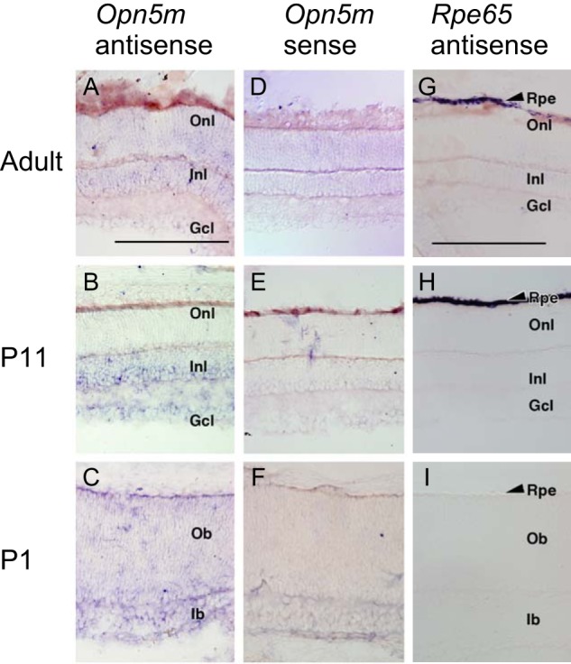
Expression of Opn5m and Rpe65 mRNA in the mouse retina. A–F, detection of Opn5m mRNA in the retina of adult (ICR strain, 8 weeks, male), P11, and P1 mice. D–F, sense probes gave essentially no signals. G–I, detection of Rpe65 mRNA in the RPE of adult, P11, and P1 mouse. I, Rpe65 mRNA was undetectable in P1 retina. Rpe, retinal pigment epithelium; Onl, outer nuclear layer; Inl, inner nuclear layer; Gcl, ganglion cell layer; Ob, outer neuroblastic layer; Ib, inner neuroblastic layer. Scale bars, 0.2 mm in A–F and 0.2 mm in G–I.