
FIGURE 9.
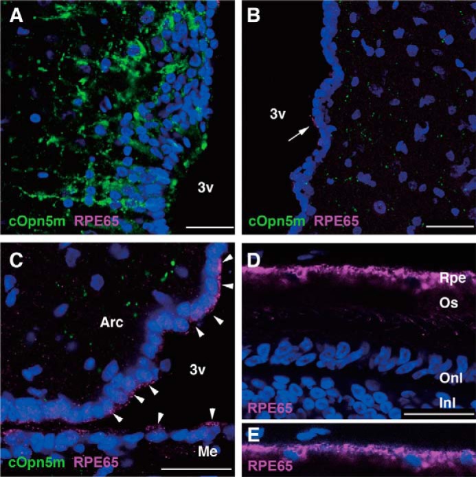
Localization of RPE65-immunoreactive cells in the chick brain. A, Opn5m-expressing neurons in the PVO. There were no RPE65-immunoreactive cells in and near the PVO. B, in the anterior hypothalamus, a subset of ependymal cells were immunoreactive for RPE65. The arrow shows RPE65 protein localized to the apical side of an ependymal cell. C, more intense expression of RPE65 protein was observed in the ependymal cells lining the ventral region of the third ventricle (arrowheads). D, the anti-RPE65 antibody recognized retinal pigment epithelium (Rpe) in the retina at P10. E, to better visualize nuclei of RPE cells, the blue channel is enhanced. Nuclei are stained with DAPI. 3v, third ventricle; Arc, arcuate nucleus; Me, median eminence; Os, outer segments of photoreceptors; Onl, outer nuclear layer; Inl, inner nuclear layer. Scale bar, 25 μm.