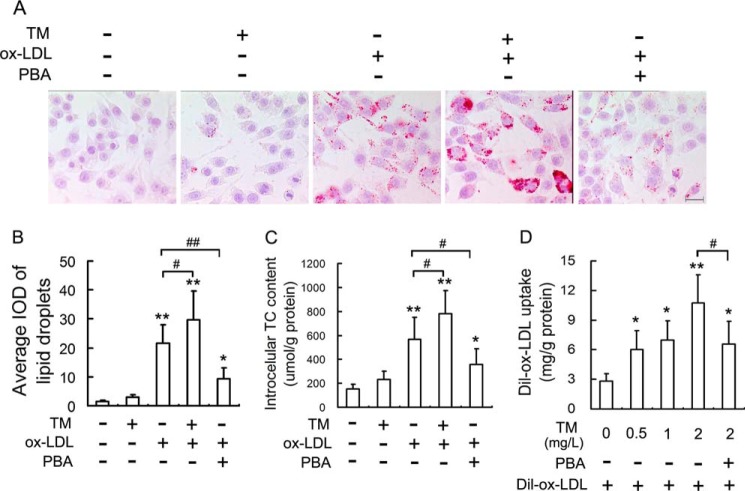
FIGURE 1.
ER stress mediates ox-LDL-induced lipid accumulation in macrophages. A, RAW264.7 cells were treated with 50 mg/liter ox-LDL in the presence or absence of tunicamycin (TM) (0.5 mg/liter) and 4-phenylbutyric acid (PBA) (20 mm) for 12 h, and the extent of lipid loading was assessed by oil red O staining. Representative lipid droplet staining images are shown. Scale bar, 20 μm. B, the average integrated optical density (IOD) of lipid droplets stained with oil red O from differentiated macrophages. C, under the same conditions as in A, the intracellular total cholesterol (TC) content was measured using a tissue/cell TC assay kit. D, Dil-ox-LDL fluorescence intensity in cells incubated with TM at the indicated concentrations in the presence or absence of PBA (20 mm) for 4 h, followed by incubation with Dil-labeled ox-LDL (50 mg/liter) for 4 h. Data are presented as the mean ± S.E. of at least four independent experiments. *, p < 0.05, **, p < 0.01 versus vehicle-treated control, #, p < 0.05, ##, p < 0.01.

