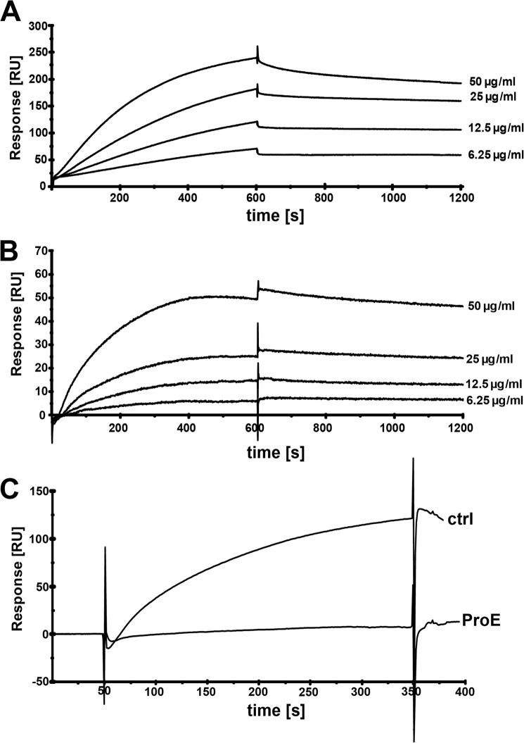
FIGURE 3.
Binding of S. aureus surface proteins to hTSP-1 as analyzed by surface plasmon resonance. A and B, surface plasmon resonance sensorgrams demonstrating a dose-dependent binding of surface proteins enriched from S. epidermidis RP62A (A) and S. aureus SA113 (B) to hTSP-1 immobilized on a CM5 biosensor. C, binding of surface proteins of S. aureus SA113 (100 μg/ml) to immobilized hTSP-1 before (ctrl) and after incubation with Pronase E (ProE). The CM5 biosensor was coated with hTSP-1 (∼7500 RU), and enriched surface proteins were used as analytes at a flow rate of 10 μl/min. The affinity surface was regenerated between subsequent sample injections with 12.5 mm sodium hydroxide. The values of the control flow cell were subtracted from each sensorgram.

