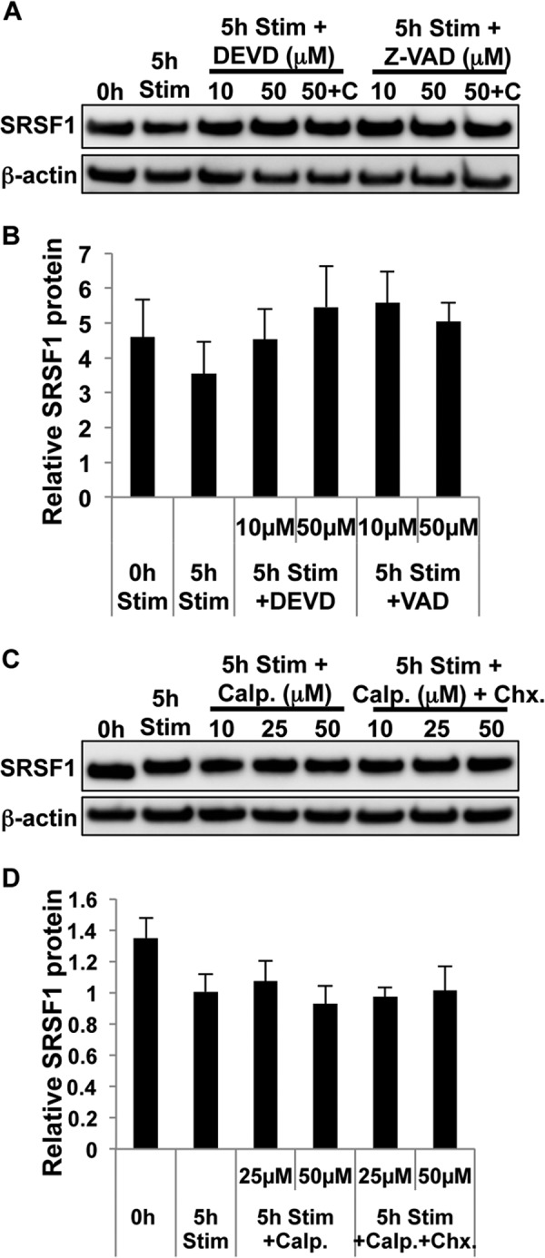
FIGURE 8.

T cell activation induces caspase-mediated degradation of SRSF1. A, T cells were pretreated with the protein translation inhibitor cycloheximide (C, 10 μg/ml) for 10 min followed by either caspase-3 inhibitor DEVD or pan-caspase inhibitor VAD for 1 h and then activated with soluble anti-CD3 and anti-CD28 antibodies for 5 h. Total protein was extracted from T cells and immunoblotted for SRSF1 and β-actin. 5h Stim, 5-h stimulation. B, densitometric quantitation of SRSF1 immunoblots from A was performed and normalized to β-actin. C, T cells were pretreated with cycloheximide (Chx, 10 μg/ml) for 10 min followed by protease inhibitor calpeptin (Calp.) for 1 h, and then activated with soluble anti-CD3 and anti-CD28 antibodies for 5 h. Total protein was extracted from T cells and immunoblotted for SRSF1 and β-actin. D, densitometric quantitation of SRSF1 immunoblots from C was performed and normalized to β-actin. Graphs show average values from n = 4 donors, and error bars represent S.E.
