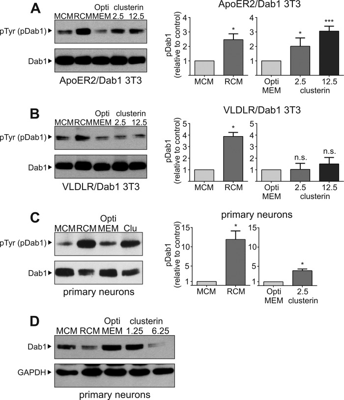
FIGURE 3.
Clusterin induces Dab1 phosphorylation and degradation. A, 3T3 cells expressing ApoER2 and Dab1 (ApoER2/Dab1 3T3), (B) VLDLR and Dab1 (VLDLR/Dab1 3T3), and (C) primary rat E16.5 WT neurons were incubated with mock-conditioned medium (MCM), Reelin-conditioned medium (RCM), OptiMEM or clusterin (A and B: 2.5 nm and 12.5 nm clusterin; C: 2.5 nm clusterin) Cells were processed for immunoprecipitation of Dab1. Anti-Dab1 antibody was used to detect total Dab1 levels, and anti-phosphotyrosine (pTyr) antibody was used to detect tyrosine-phosphorylated Dab1 (pDab1). Western blots from three independent experiments per cell line were quantified by densitometry using ImageJ 1.48 and normalized with total Dab1 levels (n = 3; plots show mean ± S.E.; n.s. not significant; *, p < 0.05 versus control; ***, p < 0.001 versus control; unpaired Student's t test was performed in GraphPad Prism 6). D, primary rat E16.5 WT neurons were incubated with MCM (lane 1), RCM (lane 2), OptiMEM (lane 3), 1.25 nm clusterin (lane 4), or 6.25 nm clusterin (lane 5) for 5 h. Total cell extracts were prepared, and Western blotting was performed using anti-Dab1 and anti-GAPDH antibodies. Experiments were repeated three times with similar results.

