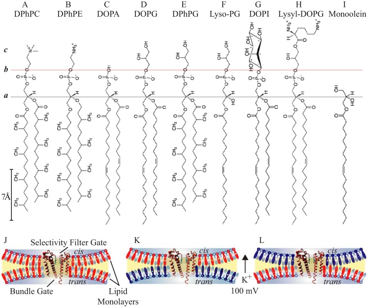
FIGURE 1.
Chemical structures of lipid molecules and bilayer configurations. Horizontal black line designates the common acyl-glycerol esterification site in all the lipids (plane a). Horizontal red line indicates a shared negatively charged phospho-X headgroup boundary, where X bears distinct head group chemistry (plane b): A, phosphocholine; B, phosphoethanolamine; C, phosphatidic acid; D–F, phosphoglycerol; G, phosphoinositol; H, lysyl-phosphoglycerol. c, denotes a layer with interfacial properties generated by the specific headgroup chemistry. I–K, schematic representation of the PM (orange) in a lipid bilayer (blue contour) depicting the orientation of the selectivity filter and bundle gates of the PM in symmetric (I) and asymmetric (J and K) bilayers. DPhPC is depicted in cyan, doping lipids are in red and dark blue, irrespective of charge.

