FIGURE 9.
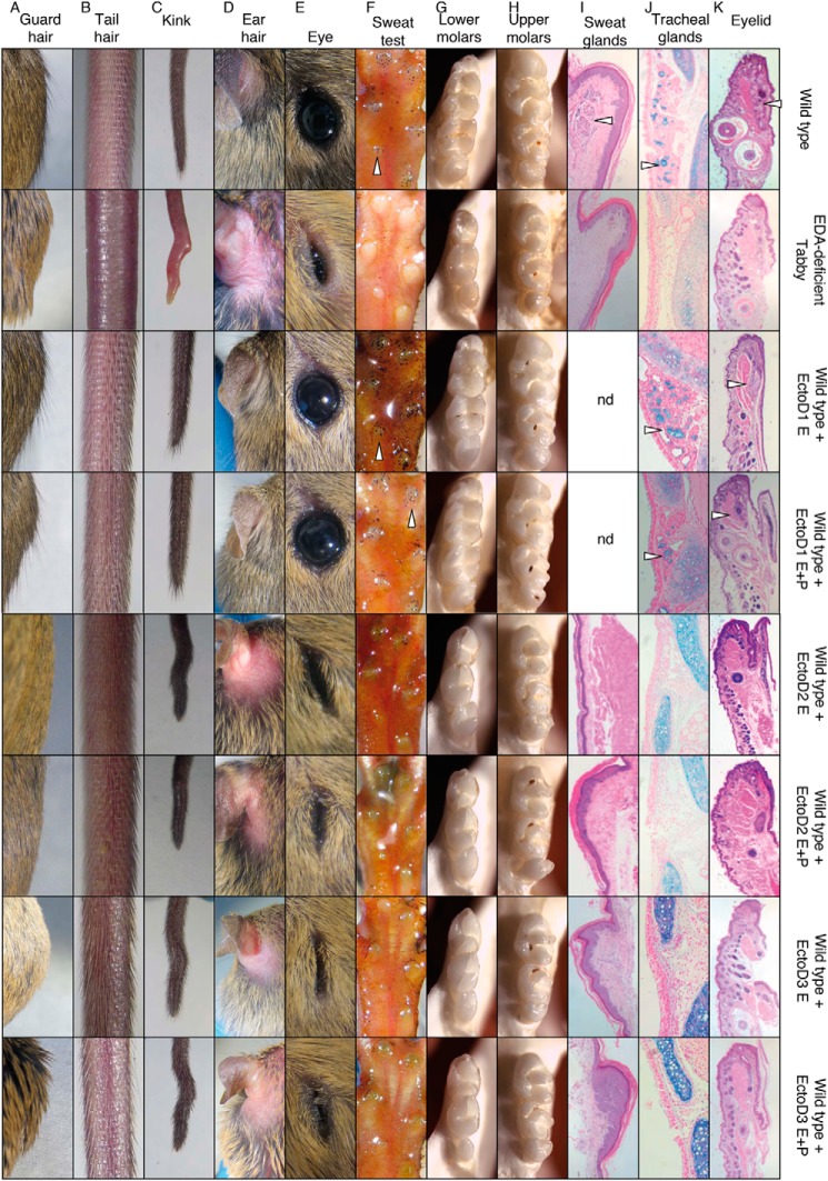
Anti-EDA monoclonal antibodies EctoD2 and EctoD3, but not EctoD1, induce ectodermal dysplasia when administered during embryonic development in wild type mice. Pregnant wild type mice were treated intravenously every second or fourth day with EctoD1 (E0, E2, E4, E6, E8, E10, E12, E14, E16, and E18 ±[P1, P2, P4, P6, P8, P10, P14, P16, and P18]), EctoD2 (E5, E9, E12, and E16 ±[P2, P6, P10, P14, and P16]), or EctoD3 (E3, E5, E8, E10, E12, E14, E16, and E18 ±[P1, P3, P5, P7, P9, P11, P13, P15, P17, and P19]) at 4 mg/kg throughout pregnancy. Treatment was stopped at birth in half of the pups and continued in the other half of the pups at the same dose up to the age of 16–19 days. All pups were analyzed at 3 weeks of age for their external appearance and at 1 month for necropsy. Untreated wild type and Eda-deficient Tabby mice were similarly analyzed for comparison. The figure shows results for one animal per group. Phenotype within a group was very similar or identical. Treatment E+P, treatment was performed during embryogenesis and continued postbirth by intraperitoneal injections in pups. Treatment E, treatment was performed during the embryogenesis only, and pups did not receive intraperitoneal injections postbirth. A, back hair with protruding guard hairs. B, tail hair phenotype. C, phenotype of the tip of the tail. D, top view of the retroauricular region. E, eye phenotype. F, footpads with functional sweat glands visualized as black dots (arrowheads) with the starch iodine test. G, lower molars, with the anterior part on the top. H, upper molars, with the anterior part on the top. I, sections of footpads with glandular structures of sweat glands (arrowhead), stained with hematoxylin and eosin. J, sections of the trachea stained with Alcian blue to reveal mucus-secreting glands (arrowheads). K, eyelid sections stained with hematoxylin and eosin, with glandular structure of meibomian glands (arrowheads).

