FIGURE 2.

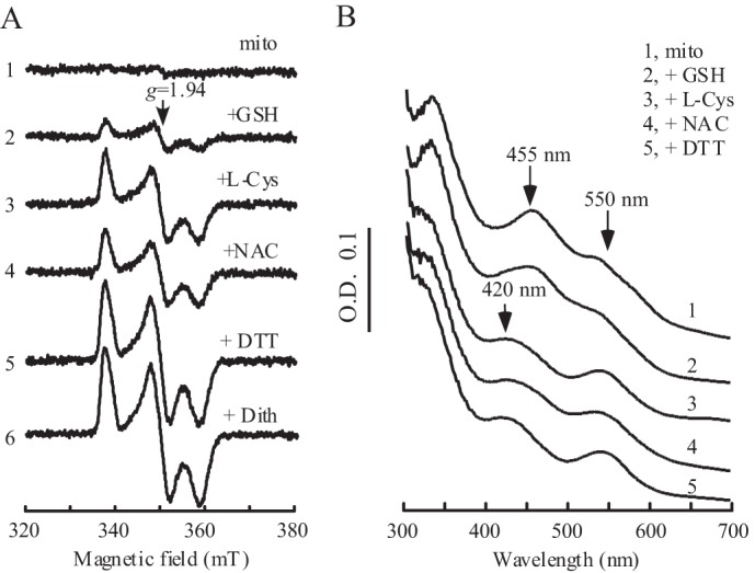
MitoNEET [2Fe-2S] clusters can be reduced by biological thiols in vitro. A, EPR spectra of the mitoNEET [2Fe-2S] clusters after reduction with different thiols. Purified mitoNEET [2Fe-2S] clusters (14 μm) (spectrum 1) were incubated with 2 mm of GSH (spectrum 2), l-cysteine (l-Cys) (spectrum 3), N-acetyl-l-cysteine (NAC) (spectrum 4), or DTT (dith) (spectrum 5) at 37 °C for 20 min under anaerobic conditions. The EPR spectrum of fully reduced mitoNEET [2Fe-2S] clusters (spectrum 6) was obtained by adding sodium dithionite (2 mm). mT, millitesla. B, UV-visible spectra of mitoNEET [2Fe-2S] clusters after incubation with biological thiols. Purified mitoNEET [2Fe-2S] clusters (14 μm) (spectrum 1) was incubated with 2 mm of GSH (spectrum 2), l-cysteine (spectrum 3), N-acetyl-l-cysteine (spectrum 4), or DTT (spectrum 5) at 37 °C for 20 min under anaerobic conditions. The absorption peaks at 455 nm and 550 nm represent the oxidized mitoNEET [2Fe-2S] clusters, and the new absorption peak at 420 nm represents the reduced mitoNEET [2Fe-2S] clusters.
