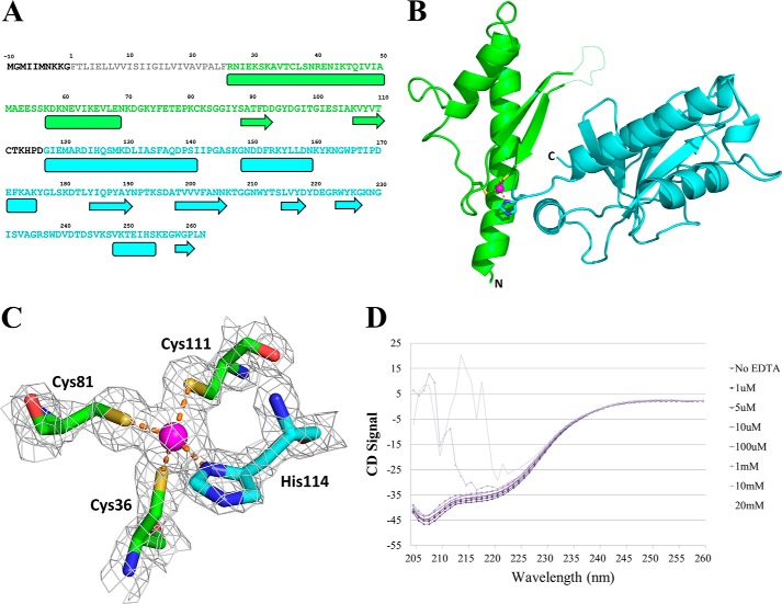
FIGURE 2.
PilJ three-dimensional structure. A, the sequence of PilJ with the secondary structure outlined below. The prepilin leader sequence is shown in black, and the α1-n region is in gray. The N-terminal domain is green, and the C-terminal domain is blue. Helices are shown as boxes, and strands are shown as arrows. B, schematic representation of the structure of PilJ. The N-terminal domain is green, the C-terminal domain is blue, and the zinc atom is magenta. The disordered loop spanning residues 94–102 is indicated with a dotted line. C, the zinc-binding site of PilJ. Cysteines 36, 81. and 111 from the N-terminal domain are in green, histidine 114 from the C-terminal domain is in cyan, and the zinc atom is in magenta. The gray mesh shows the bounds of a 2Fo − Fc electron density map. D, CD spectra of PilJ are shown in increasing concentrations of EDTA.

