
Figure 2.
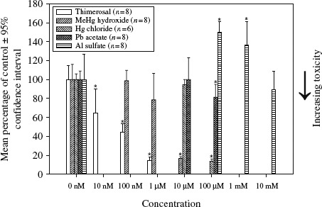
A concentration-dependent assessment of metal-induced mitochondrial dysfunction in human neuroblastoma cells following 24 h incubation.
Notes: Mitochondrial dysfunction was measured using the XTT cell assay (following 2 h incubation). *p < 0.05 (Exposure concentration in comparison with the 0 nM Control). Thimerosal LC50 = 82.2 nM, MeHg hydroxide LC50 = 5.6 µM, Hg chloride LC50 = 59.5 µM, Pb acetate LC50 > 100 µM, Al sulfate LC50 > 10 mM.