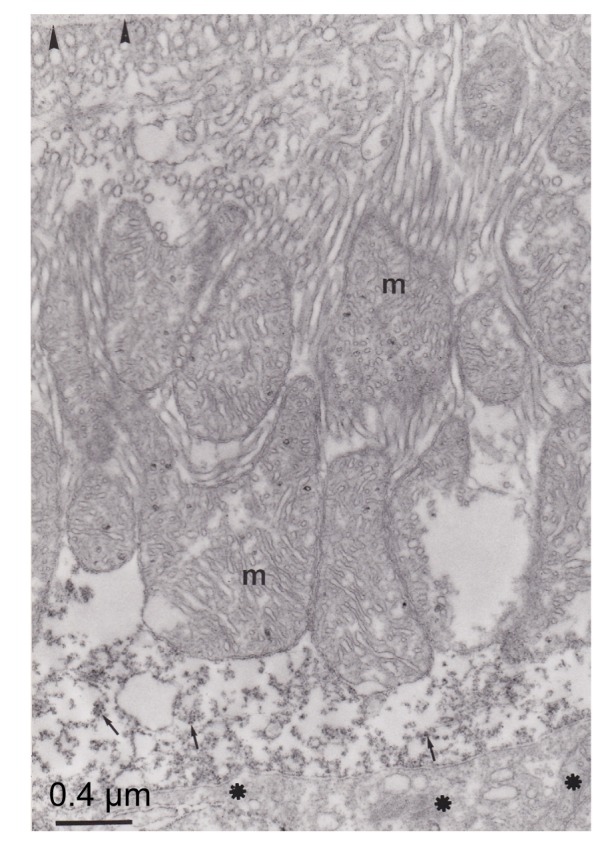
Fig. 6.

Electron micrograph of pseudobranch cells
Note that the immunoreactivity was distributed in the apical part of the cell (arrow). Lacunars tissue (asterisks) is not marked. Basement membrane (arrowhead) was not immunostained. Mitochondria (m) and tubular system are not marked
