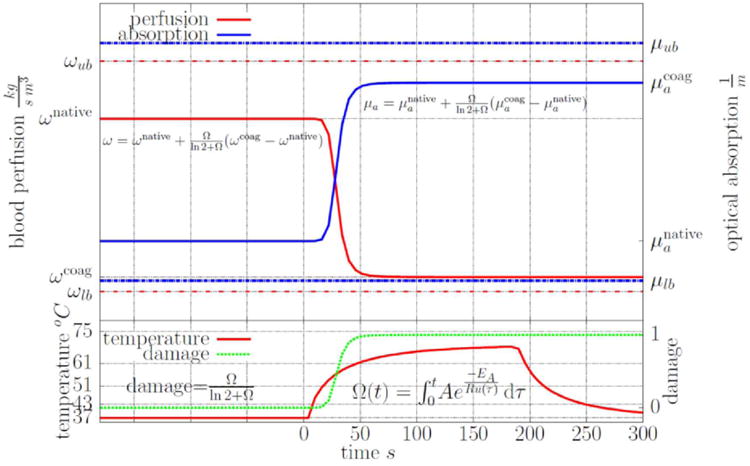
Figure 1.

A graphical illustration of the nonlinear constitutive model [27] used for the perfusion and optical absorption is shown. The scattering behaves similar to the absorption. Damage, perfusion, and absorption are plotted as a function of a time-temperature history representative of those observed in MRgLITT procedures. As shown, the native undamaged constitutive values transition to coagulated constitutive values between the temperature range of approximately 51-61 °C. Similar to previous results [49], the tissue is fully damaged at a 61 °C threshold.
