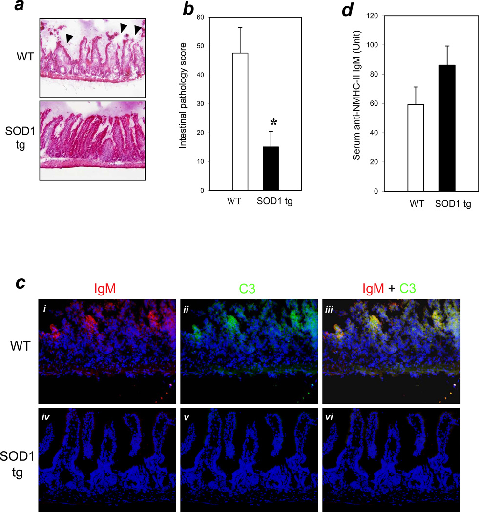
Figure 1. SOD1 overexpression decreased tissue injury and blocked IgM-mediated complement activation in the intestinal I/R model.
(a) Intestinal I/R (40 min./3 hrs.) was carried out and H&E staining performed as described in Methods. Arrows indicate typical pathology features of villi injury (200× magnification). (b) Bar graph: bars indicate mean pathology scores based on the degree of villi injury as described in Methods. Error bars indicate standard error of the mean (SEM). Asterisks indicate statistical significance (P<0.01; n=12 for WT, n=11 for SOD1 tg). (c) SOD1 overexpression results in the blocking of IgM and C3 binding in vivo. Panels i–iii: WT mice; panels iv-vi: SOD1 tg mice. Representative cryosections were stained with anti-IgM-biotin/streptavidin-Alexa-568 (red fluorescence in panels i and iv) and anti-C3-FITC (green fluorescence in panels ii and v). Sections were counterstained with DAPI (Violet/Blue). Panels iii and vi are merged images of (i + ii) and iv + v) respectively. Magnification=400×. (d) Serum levels of anti-NMHC-II IgM were similar in SOD1 tg and WT mice (n=5 for each group; P>0.05 between these groups).

