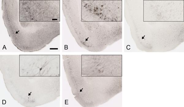
Figure 4. Anti-tau antibodies strongly decreased AT8 staining in P301S mouse brain.
Representative coronal sections of PBS (A), HJ3.4 antibody (B), HJ8.5 antibody (C), HJ9.3 antibody (D) and HJ9.4 antibody (E) treated 9 month old P301S mice stained with biotinylated AT8 antibody in regions including the piriform cortex and amygdala. Scale bar is 250 μm. Inserts in A to E show the higher magnification of biotinylated AT8 antibody staining of phosphorylated tau, scale bar is 50 μm.

