Fig. 4.

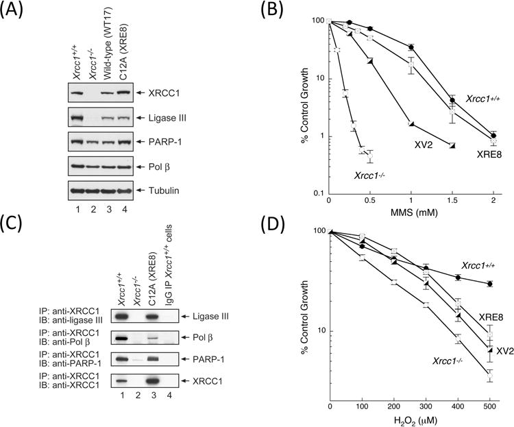
Characterization of XRE8 cells expressing disulfide blocked reduced XRCC1 protein. (A) Western blotting analysis of XRCC1 and other repair proteins in XRE8 cells compared with Xrcc1+/+, Xrcc1−/− and WT17. (B) Comparison of complementation of MMS hypersensitivity of Xrcc1−/− cells by stable expression of C12A (XRE8 cells) or V88R protein (XV2 cells). Full methods for growth inhibition assays are described in Section 2. (C) Equal amounts of cell extracts as indicated were immunoprecipitated with anti-XRCC1 antibody then immunoblotted with ligase III, pol β, PARP-1 and XRCC1 antibodies. In lane 4, immunoprecipitation was of Xrcc1+/+ cells with pre-immune IgG as a negative control. Full methods are described in Section 2. (D) Comparison of complementation of H2O2 hypersensitivity of Xrcc1−/− cells in XRE8 cells or XV2 cells measured by growth inhibition assay.
