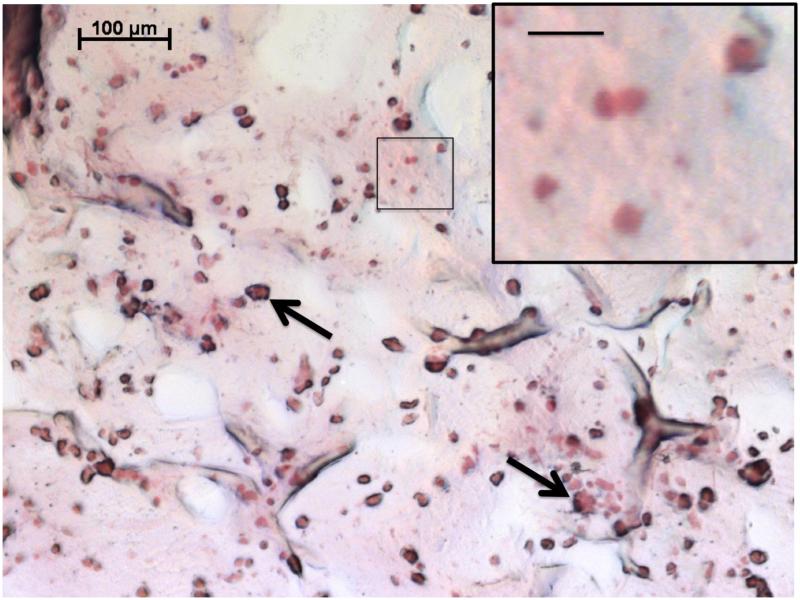
Figure 6.
Representative histological section of the chondral layer of cell-laden OPF hydrogel composites stained with Safranin-O at Day 28. The higher magnification image depicts the spherical morphology of encapsulated cells, where the scale bar represents 20 μm. Arrows indicate gelatin microparticles, now partially degraded, that were also encapsulated within the construct. This section was taken from Group 2 (MSC/OS), which is representative of histology from all groups examined.

