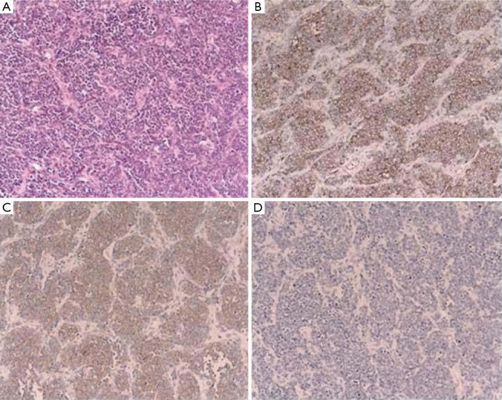
Figure 2.
Pathology and Immunohistochemical staining of the tumor. A. Hematoxylin and eosin stain (original magnification ×150) of a PNET (primitive neuroectodermal tumor) of the pancreas. These neoplasms were composed of sheets of small round cells with hyperchromatic round to oval nuclei. The cells had no Homer-Wright rosettes; B. Immunohistochemical stain (original magnification ×150) for CD99 (p30/32MIC2). The neoplastic cells showed strong cytoplasmic membrane positive to the MIC-2 glycoprotein; C. Immunohistochemical stain (original magnification ×150) for neuron-specific enolase showing strong focal positive and demonstrating the neuronal differentiation of these small round cell tumors; D. Immunohistochemical stain (original magnification ×150) for cytokeratin (AE1/AE3) showing negative

