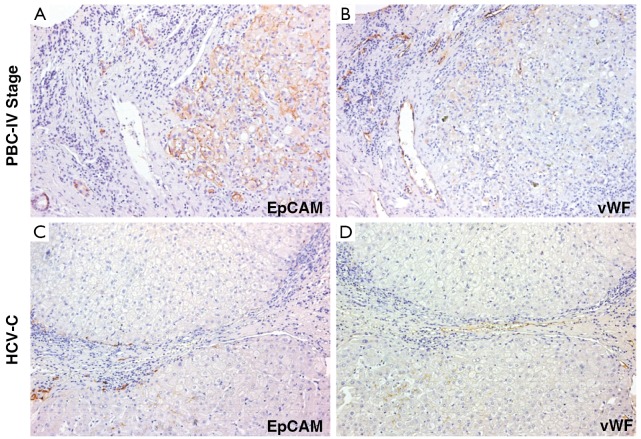
Figure 1.
Immunohistochemistry for EpCAM and von Willebrand Factor (vWF) in serial sections of liver biopsies of Primary Biliary Cirrhosis (PBC) and HCV-related cirrhosis (HCV-C) patients. (A) PBC samples show a more extensive expansion of HPC population (EpCAM-positive cells) compared with those of HCV-C samples (C). In PBC, progenitor cells penetrate into cirrhotic nodules and several EpCAM+ hepatocytes (newly derived from HPCs) could be observed; in HCV-C samples, reactive ductules are located within the fibrous septa and at the interface with cirrhotic nodule; (B) PBC samples show a more extensive angiogenesis (vWF-positive endothelial cells) if compared to HCV-C biopsies (D). For semi-quantitative analysis see Table 1. Original magnification 20× (A-D)

