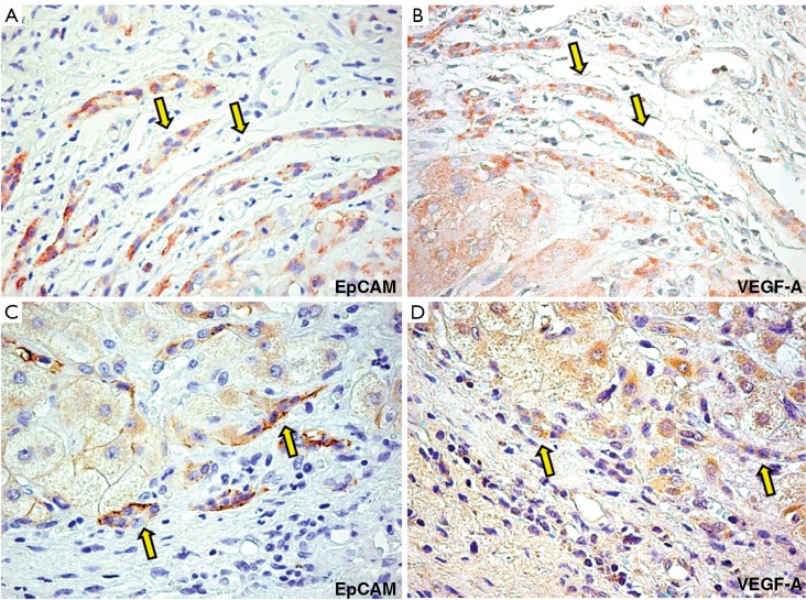
Figure 4.
Immunohistochemistry for EpCAM (A) and VEGF-A (B) and EpCAM (C) and VEGF-C (D) in serial sections of PBC biopsies. PBC samples are characterized by an increased expression of VEGF-A (yellow arrows) and VEGF-C (yellow arrows). For semi-quantitative analysis see Table 1. Original magnification 40× (A-D)

