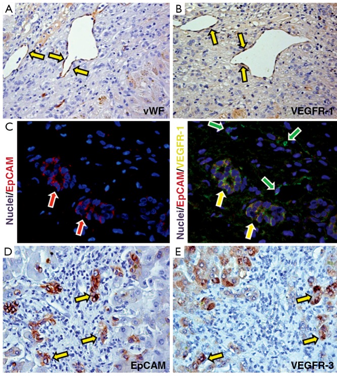
Figure 5.
(A,B) Immunohistochemistry for von Willebrand Factor (vWF: A) and VEGFR-1 (B) in primary biliary cirrhosis (PBC) samples. In PBC, endothelial cells (A) express VEGFR-1 (B); (C) Double immunofluorescence for Epcam (red channel) and VEGFR-1 (green channel); nuclei are visualized in blue channel in PBC biopsies. Numerous EpCAM-positive progenitor cells co-express VEGFR-1 (yellow cells: yellow arrows). Notably, endothelial cells in vessels are positive for VEGFR-1 (green arrows); (D,E) Immunohistochemistry for EpCAM (D) and VEGFR-3 (E) in serial sections of PBC biopsies. EpCAM positive HPCs highly express VEGFR-3 (arrows)

