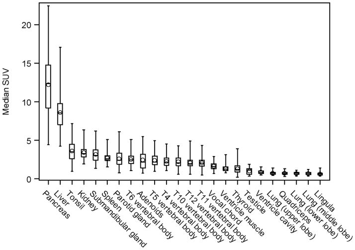
FIGURE 1.
The SUVs for the tissues examined. Individual data points are depicted, along with the group median (solid horizontal line in the rectangular box), the group mean (◇ symbol), the first and third quartiles above and below the group median (rectangular box), and the minimum and maximum SUV values (top and bottom horizontal lines.

